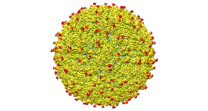
Zika structure mapped for first time

Zika now has a face to go with the name.
New microscopy images of the virus reveal a bumpy, golf ball‒shaped structure, similar to that of the dengue and West Nile viruses, researchers report March 31 in Science. It’s the first time scientists have gotten a good look at Zika, the infamous virus that has invaded the Americas and stoked fears that it is causing birth defects and a rare autoimmune disease (SN: 4/2/16, p. 26).
Cracking Zika’s structure is like getting the blueprints of an enemy’s base: Now scientists have a better idea of where to attack. “This certainly gives us great hope that we will be able to find a vaccine or antiviral compounds,” says study coauthor Michael Rossmann of Purdue University in West Lafayette, Ind., who’s known for mapping the first structure of a common cold virus in 1985.
Researchers have been racing to solve Zika’s structure, says UCLA microbiologist Hong Zhou. “I was trying to work on the same thing myself.” But the new study’s authors beat everybody. “I was impressed they were able to do it so quickly,” Zhou says.
Rossmann and colleagues imaged a strain of Zika collected from a patient during a 2013‒2014 outbreak in French Polynesia (the strain is nearly identical to the one now spreading through Latin America).
The team used a technique called cryoelectron microscopy to create a three-dimensional picture of Zika. It’s a pretty sharp image, says study coauthor Devika Sirohi, also of Purdue. She and colleagues can clearly see the virus’ shape and can even make out sugars protruding from its surface.
These sugars, which look like little red doorknobs, hang from proteins in Zika’s shell. The knobs may help Zika attach to — and infect — human cells. The team discovered that Zika’s knobby regions look slightly different from those of related viruses. Zika’s sugar-decorated proteins “fold a little differently,” Sirohi says. And that might let Zika make different contacts with attachment sites on cells, called receptors. That could “influence what kind of cell the virus infects,” she says. These differences could explain why Zika infects cells not typically targeted by dengue or West Nile.
One of the receptors targeted by Zika could be AXL, a protein crowded on the surface of neural stem cells, researchers propose March 30 in a separate study published online in Cell Stem Cell. Zika virus is thought to preferentially infect these early-development brain cells, and it could potentially use AXL as an easy entry point, study coauthor Arnold Kriegstein of the University of California, San Francisco and colleagues suggest.
Of course, exactly what role subtle structural differences play in Zika’s infection ability is “only speculation at this point,” Sirohi says. The team now plans to test how tweaking the knobby regions of the virus affects Zika’s virulence.